StarCare Animals
Cats and dogs that develop urinary tract obstruction, such as urinary stones, urethral obstruction, or tumors, may cause significant systemic effects, including changes on the electrocardiogram (ECG). These changes are usually associated with electrolyte imbalances, particularly hyperkalemia, which is an abnormally high level of potassium in the blood.
Hyperkalemia due to urinary tract obstruction may cause the following ECG changes:
1. Spiking T waves: one of the earliest changes to appear on the ECG with elevated blood potassium levels are spiking T waves; these T waves are usually narrow and symmetrical.
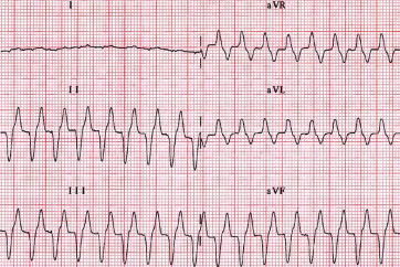
2. Prolongation of the PR interval and QRS complex: as potassium levels rise further, the conduction system may be delayed as evidenced by first a prolongation of the PR interval followed by a widening of the QRS complex.
3. Disappearance of P-waves: at higher potassium levels, P-waves may become flattened or even disappear completely, making atrial activity difficult to recognize on the ECG.
4. Sine wave pattern: In severe hyperkalemia, the ECG may show a sinusoidal wave pattern, which is a serious indicator of severely elevated blood potassium and may lead to death if left untreated.
5. Bradycardia and other arrhythmias: High potassium levels may also lead to a variety of cardiac arrhythmias, including bradycardia (slow heartbeat), ventricular fibrillation, or cardiac arrest.
6. Atrial Arrest: In severe cases of hyperkalemia, atrial arrest, a phenomenon in which atrial activity cannot be observed on the electrocardiogram (ECG), may occur. This is a serious condition that can potentially lead to life-threatening arrhythmias. In this condition, the normal rhythmic regulation of the heart is impaired and the atria are unable to send electrical signals properly, thus affecting the function of the entire heart and blood circulation. This cardiac condition requires urgent medical intervention to prevent further cardiac complications and potentially fatal outcomes.
The severity of these ECG changes is closely related to the level of hyperkalemia. Therefore, close monitoring and management of electrolyte imbalances in these animals is critical to prevent potentially life-threatening cardiac complications. Rapid veterinary intervention to relieve urinary tract obstruction and correct electrolyte imbalances is critical to the survival of the animals.
References
Scott A. Brown, Obstructive Uropathy in Small Animals, https://www.merckvetmanual.com/urinary-system/noninfectious-diseases-of-the-urinary-system-in-small-animals/obstructive-uropathy-in-small-animals#Treatment:_v3295921, Oct 2013
Amy Butler, Feline Urethral Obstruction, https://www.delawarevalleyacademyvm.org/pdfs/jun14/FullNotes.pdf
Kelly St. Denis, Managing Feline Urethral Obstruction, https://www.todaysveterinarypractice.com/urology-renal-medicine/managing-feline-urethral-obstruction/, Sept 2020
Teresa M. Rieser, Urinary Tract Emergencies, Vet Clin Small Anim, 35 (2005) 359–373

Urethral Obstruction in Cats (https://www.cliniciansbrief.com/article/urethral-obstruction-cats).

Hyperkalemia in the dog – T waves become tall and spiked (https://obivet.com/lessons/hyperkalemia/).


Wide-complex tachycardia and absent P waves. Serum potassium=9.2 mEq/l (https://doi.org/10.1016/j.jfms.2006.04.003).