StarCare Animals
Specific ECG Changes in Valvular Heart Disease:
|
Valvular heart disease (VHD), particularly degenerative mitral valve disease (DMVD), is common in dogs. DMVD is the most prevalent cardiac condition and a leading cause of congestive heart failure in canines. The incidence of VHD in small breed dogs increases significantly with age.
Degenerative Valvular Disease (DVD) affects about 85% of elderly small dogs and typically progresses over several years. The mitral valve is most frequently affected, though the tricuspid valve is also involved in approximately 30% of cases. This condition is chronic and progresses slowly, often lasting over five years and ultimately leading to heart failure and death. Notably, male dogs are more susceptible than females.
The main pathological change of DVD is myxomatous degeneration, where the valve leaflets lose collagen and other connective tissues while abnormal accumulations of proteoglycans occur. These changes lead to a loss of the valve’s normal supporting structure and leaflet deformation, resulting in valvular regurgitation. As the condition worsens, the forward cardiac output decreases, triggering various compensatory mechanisms to meet the body’s circulatory demands.
Diagnosing DMVD is usually straightforward, primarily based on the presence of a left-sided systolic murmur in small adult dogs. As the disease progresses, thoracic radiographs may reveal an enlarged cardiac silhouette, especially of the left atrium and ventricle. ECG may show changes consistent with left ventricular or biventricular enlargement, particularly in advanced stages of the disease.
In dogs with myxomatous mitral valve disease (MMVD), changes in ECG parameters significantly correlate with disease progression. A study published on PubMed evaluated these changes and their predictive value for congestive heart failure (CHF). Results showed that as mitral valve disease progresses from stage B to stage C, there is a significant prolongation of P wave duration and corrected QT interval (QTc). Specifically, dogs with stage C MMVD had significantly longer P wave duration and QT interval compared to those in stage B. These findings indicate that the prolongation of P wave duration and QTc in MMVD dogs can help predict the onset of CHF.
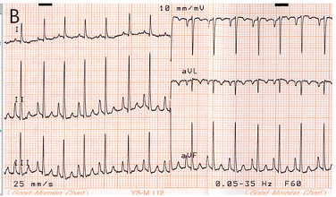
Additionally, a study by IntegOpen explored the various physical and ECG changes caused by chronic mitral valve insufficiency (CMVI) in dogs. One notable ECG finding in dogs with CMVI is the so-called "P mitrale," characterized by wide P waves and tall QRS complexes, indicating left atrial (LA) and left ventricular (LV) enlargement. These ECG changes are crucial for diagnosing and assessing the severity of the condition. The study also highlighted the importance of physical examination, laboratory tests, cardiac biomarkers, and echocardiography in the evaluation and management of canine heart disease.
Overall, ECG changes in dogs with valvular heart disease, especially MMVD and CMVI, are significant and provide valuable information about disease progression and severity. Key observed changes include the prolongation of P wave duration and QTc interval, which help predict CHF. Additionally, the presence of "P mitrale" and changes in the QRS complex indicate LA and LV enlargement in dogs with CMVI, essential for diagnosis and management.
The treatment goals for DMVD are to alleviate symptoms, control congestion and edema, and slow disease progression. For dogs with advanced mitral valve insufficiency, appropriate treatment, including diuretics, ACE inhibitors, and inotropic agents like pimobendan, can help maintain quality of life, potentially extending it by months to years. Although surgical valve replacement or repair is becoming more common in veterinary practice, its use is still limited due to cost and equipment constraints.
References
Na Y, etc., Comparison of electrocardiographic parameters in dogs with different stages of myxomatous mitral valve disease, Can J Vet Res. 2021 Oct; 85(4):261-270.
Suh S, etc., “Chronic Mitral Valve Insufficiency in Dogs: Recent Advances in Diagnosis and Treatment”, in Canine Medicine - Recent Topics and Advanced Research edited by Hussein Abdelhay Elsayed Kaoud, December 2016, DOI: 10.5772/65689
Pinkos A and Stauthammer C, Degenerative Valve Disease: Classification, Diagnosis, and Treatment of Mitral Regurgitation, Today’s Veterinary Practice, September/October 2021
Oyama M, An Everyday Approach to Canine Degenerative Mitral Valve Disease, Today’s Veterinary Practice, July/August 2012

Mitral valve disease in dogs (https://www.pdsa.org.uk/pet-help-and-advice/pet-health-hub/conditions/mitral-valve-disease-in-dogs).

ECG in dogs with CMVI. P‐mitrale (wide P‐wave) and wide and tall QRS complexes indicating LA and LV dilation (https://www.researchgate.net/figure/A-Phonocardiogram-in-dogs-with-CMVI-Heart-murmur-is-gradually-radiated-and-louder-and_fig1_312426528).